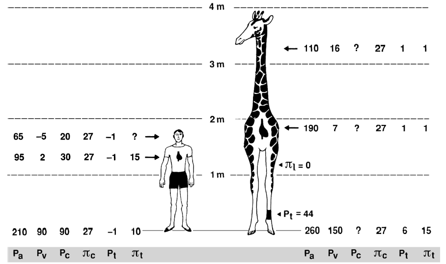
Though a giraffe’s heart is larger than that of many other animals – it’s 0.6 meters (2 feet) long and weighs about 11 kilograms – the great height of a giraffe still makes it hard for the heart to pump blood to the brain. This problem is overcome by a series of one-way valves that force blood toward the head. Giraffes are also able to put plenty of oxygen into their blood because they have tremendous lungs – they can hold 55 liters of air. Studies by Goetz, Pattersson, Van Citters, Warren and their colleagues revealed that arterial pressure near the giraffe heart is about twice that in humans, to provide more normal blood pressure and perfusion to the brain.
The giraffe has an extremely high blood pressure (280/180 mm Hg), which is, as said before, twice that found in humans. Additionally, the heart beats up to 170 times per minute. That is double the heart beat of humans. It was previously thought that a giraffe had a really big heart, but recent research has revealed that there isn’t room in the body cavity for this. Instead, the giraffe has a relatively small heart (relative to the size of the animal) and its power comes from a very strong beat as a result of the incredibly thick walls of the left ventricle.
The right ventricle pumps the blood a short distance to the lungs, and the muscle is about 1 cm thick. The left ventricle has to pump the blood all the way up to the head against the hydrostatic pressure of the blood already in the long vertical artery. A giraffe’s heart has evolved to have thick muscle walls and a small radius giving it great power to overcome this pressure.

}
The thickness of the muscle wall is related almost directly to the length of the neck. For every 15 cm increase in the length of the neck the left ventricle wall adds another 0.5 cm thickness.
During the evolution, the stroke volume became increasingly constrained by concentric eutrophy of the ventricle in response to the progressive evolution of a longer neck (i.e. increasing the vertical distance between the heart and head and thus requiring higher mean arterial pressure), the requirements on oxygen delivery during locomotion were probably alleviated, as the legs also got longer.
In recent years, however, some interesting studies have been carried out comparing the heart of giraffes, always considered with exceptional characteristics, and that of other mammals. A study published in the Journal of Experimental Biology in 2016 is particularly important.
Based on their tracking of the cardiomyocytes in the giraffe left ventricle, the researchers explain that there does not appear to be any significant principal difference in the myocardial architecture between the giraffe and other mammals. Therefore, there is no indication that giraffes have evolved cardiomyocytes generating excessive force or other peculiar adaptations; instead, the ability to develop a sufficiently high pressure for cerebral perfusion in the standing position can merely be ascribed to the thick myocardial wall and the small radius. Some findings carried out by the researchers could indicate that the left ventricles of the giraffe have a slightly altered myocardial architecture that can serve to redistribute the myocardial stresses differently during cardiac contraction than the myocardium of other species, which have a thickness of the ventricular wall lower than the cavity diameter ratio. The systemic vascular resistance (SVR) of giraffes is considerably higher than that of other similar-sized mammals. Given the low cardiac output, the high SVR is required to support the high mean Pa necessary to perfuse the brain. One aspect that still needs to be investigated is insight into the specific mechanisms underlying the high resistance.
Another interesting question is how giraffes avoid pooling of blood and tissue fluid (edema) in dependent tissue of the extremities. As confirmed by studies of Professor Alan R Hargens, the blood and tissue fluid pressures that govern transcapillary exchange vary greatly with exercise. These pressures, combined with a tight skin layer, move fluid upward against gravity. Other anatomical adaptations in dependent tissues of giraffes represent developmental adjustments to high and variable gravitational forces. These include vascular wall hypertrophy, thickened capillary basement membrane and other connective tissue adaptations.
In 2016, Smerup et al. also have assumed that a hypothetical perfect left ventricle, i.e. a pumping chamber that would have achieved the high mean arterial pressure of giraffes and thus retains the beneficial ratio of radius versus wall thickness reported here while still being able to generate a cardiac output per body mass unit comparable to other mammalian species would have an end diastolic volume of 1060 ml and a stroke volume of 583 ml, which is approximately twice the measured values. The cavity radius would be 4.7 cm and the myocardial wall thickness 3.3 cm. As it is not easy to calculate the total mass of this hypothetical pump, it can only be speculated that the energy cost of such an organ would likewise exceed the average mammalian value by a factor of two or three. The difficulty in studying the heart of these animals is that some types of investigations have been carried out in recent years. The study of Smerup cited above, for example, was the first to report the use of echocardiography on the giraffe.
References:
Smerup M, et al.”The thick left ventricular wall of the giraffe heart normalises wall tension, but limits stroke volume and cardiac output “
Journal of Experimental Biology 2016 219: 457-463; doi: 10.1242/jeb.132753
Hargens A., et al. The Gravity of Giraffe physiology in CUNY666B-12 CUNY666A/Aird 0 521 85376 1 December 15, 2006 13:23